|
|
Giant Cavernous Hemangioma of the Liver
Submitted by Matt Hoffman, MD
- Hemangiomas are the most common benign hepatic neoplasm
- Incidence of approximately 20%
- Occur more commonly in women
- These lesions tend to be stable, though may enlarge during pregnancy or with estrogen administration
- Clinical findings
- Usually asymptomatic and discovered incidentally
- Large lesions may cause pain, nausea, or vomiting secondary to extrinsic compression of adjacent bowel, rupture, hemorrhage, or thrombosis
- Large hemangiomas may sequester platelets and produce thrombocytopenia and DIC, a condition know as Kasabach-Merritt syndrome
- Histopathology
- Hemangiomas are composed of large vascular channels filled with slowly moving blood and are lined by a single layer of endothelial cells
- Giant hemangiomas (measuring greater than 6 cm) can contain hemorrhage, thrombus, calcification, or fibrosis
- Larger lesions are less common, comprising less than 10% of all hemangiomas
- Imaging
- Since hemangiomas are often detected incidentally, the importance of imaging lies in the fact that they must be differentiated from more clinically significant lesions, such as primary and secondary hepatic malignancies
- Ultrasound
- Usually homogeneous
- Well-defined hyperechoic masses (though few can appear relatively hypoechoic when imaged within a fatty liver)
- Giant lesions can appear heterogeneous secondary to internal complex composition
- Posterior acoustic enhancement is commonly seen
- Nuclear Imaging
- On 99mTc-sulfur colloid scintigraphy, hemangiomas appear as focal photopenic areas
- 99mTc-pertechnetate-labeled RBC SPECT is a more specific examination
- Early dynamic images yield focal photopenic areas that gradually increase in activity over time
- Limited utility for lesions smaller than 2-3cm due to poor spatial resolution
- CT
- Focal, well-circumscribed, low attenuation lesions on pre-contrast images
- Nodular, peripheral centripetal enhancement on dynamic contrast enhanced imaging
- MRI
- Has a sensitivity and specificity of greater than 90% and is the imaging modality of choice
- Typically hemangiomas are homogeneously hypointense relative to the liver on T1-weighted and markedly hyperintense (lightbulb sign) on T2-weighted images relative to the liver
- On dynamic, contrast-enhanced MR imaging, hemangiomas can demonstrate immediate homogeneous enhancement (lesions < 1.5cm)
- Peripheral, nodular centripetal enhancement pattern progressing to homogeneity (lesions 1.5-5cm)
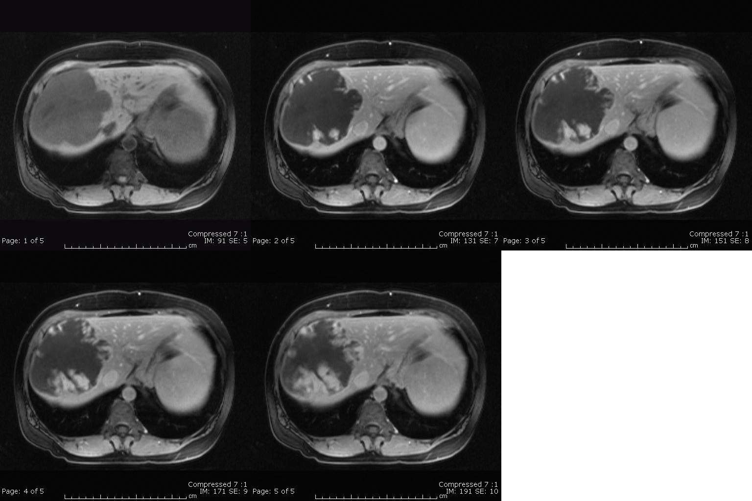
- Peripheral nodular centripetal enhancement with persistent central hypointense region (lesions> 5cm)
- Giant hemangiomas often do not achieve complete isointensity on delayed images due to their complex internal composition
- These larger lesions still should demonstrate the characteristic peripheral nodular enhancement
- MRI appearance is that of a well-defined, heterogeneous mass with areas of bright signal on T2-weighted images, cleft-like areas of low intensity on T1, and low intensity internal septae on all pulse sequences

Giant cavernous
hemangioma of the
liver: Axial T1-weighted
pre-contrast image shows a
hypointense mass
within the right
hepatic lobe.
Sequential enhanced
delayed images show
peripheral nodular
centripetal
enhancement with
persistent central
hypointensity
Semelka RC, Sofka CM:
Hepatic Hemangiomas. MRI Clinics of North
America. Volume 5 Number 2:241-252. May 1997.
|
|
|